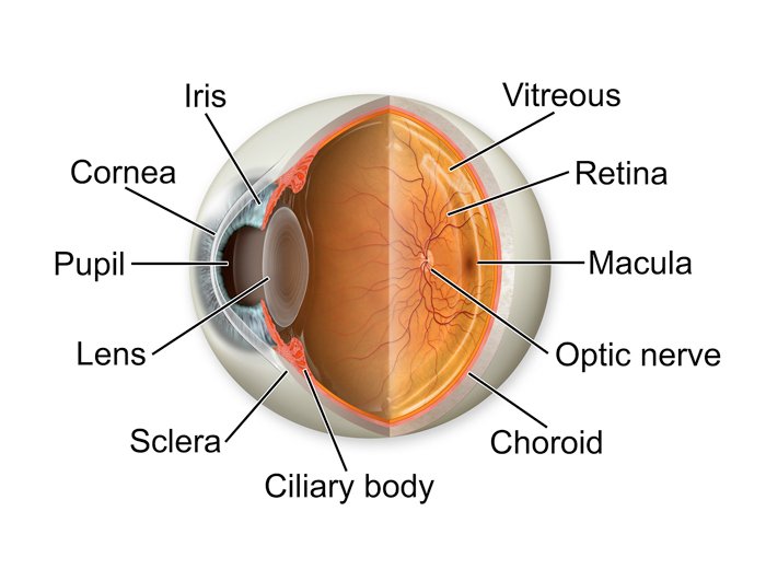
The eyeball contains three layers:
- The outer layer, formed by the cornea and sclera
- The middle layer, holding the primary blood supply for the eye and containing the iris and pupil
- The inner layer, comprised of the retina
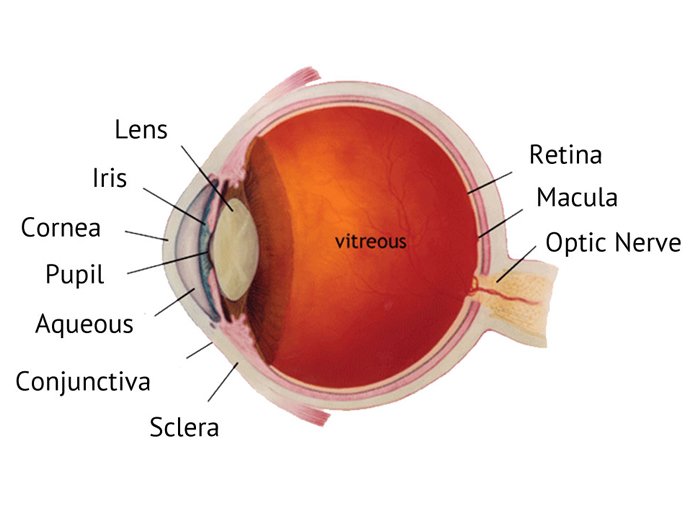
The eyeball also contains three chambers of fluid:
- Anterior chamber, between the cornea and iris
- Posterior chamber, between the iris and the lens
- Vitreous chamber, between the lens and the retina
The anterior and posterior chambers are filled with aqueous humour, which is a watery fluid that provides nourishment to the interior eye structures and helps to keep the eyeball inflated. The vitreous chamber is filled with a thicker fluid called vitreous humour, a transparent gel which is 99% water, which helps the eyes to stay inflated.
As well as numerous blood vessels, the eye also contains the optic nerve. This runs from the back of the eyeball, through an opening in the orbit known as the optic foramen.
From here, the optic nerve connects to the brain and acts as a conduit, transmitting visual information into the brain. Other nerves within the eye carry non-visual information and send messages about pain or help to control motor activity within the eye.


The cornea is the clear portion of the eye that covers the iris and the pupil and takes up about one-sixth of the eye. The rest of the eye (the scleral segment) is opaque. Several nerves and blood vessels run through the sclera, including the optic nerve. The cornea and scleral segment come together in an area called the limbus. This contains a great deal of blood vessels.
The iris and pupil are the most noticeable parts of the eye. The iris is the coloured ring of tissue that lies beneath the cornea and can be a range of colours, determined by genetics. The pupil is located in the centre of the iris and appears as a black hole that acts rather like a camera aperture, allowing light to enter the eye. This works in the same way as a camera, adjusting to control the flow of light into the eye. In bright conditions, the pupil closes down, reducing the amount of light entering the eye and protecting the delicate nerves from being damaged. In the dark, the reverse happens to allow what light there is to enter the eye.

Directly behind the iris is the lens. This focuses rays of light onto the retina, which is a light-sensitive nerve tissue that contains photosensitive cells called rods and cones. These convert light into electrical signals that are carried to the brain by the optic nerve. Every year the lens becomes slightly thicker, which is why from the age of 40 onwards it
becomes increasingly difficult to focus on near objects as the lens is too thick to flex and change shape to give a more powerful focus. Eventually this thickening process leads to cataract, this is when areas of the lens become so thick light cannot pass through them.